Research image contest highlights importance of visuals in research
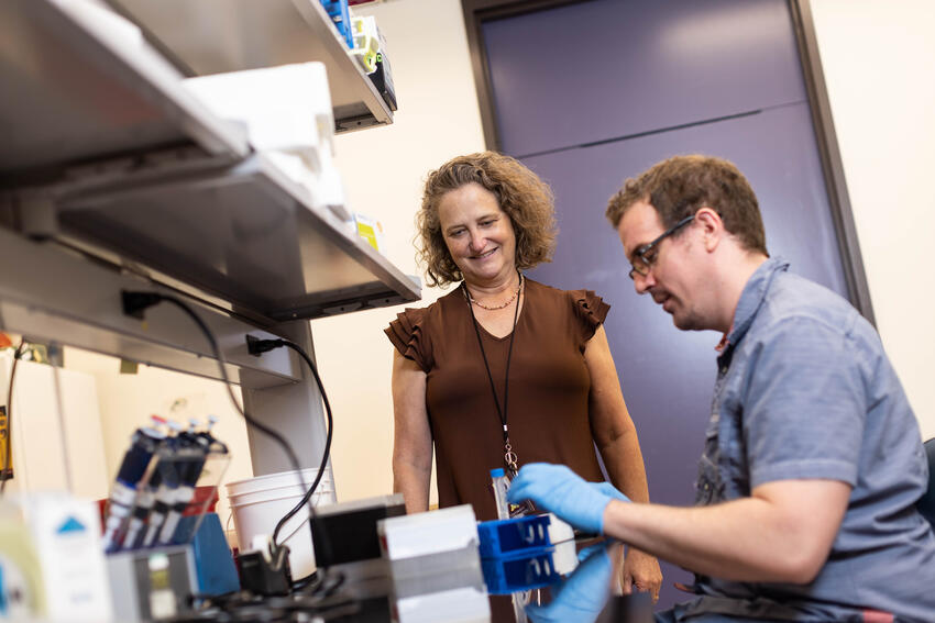
A faculty member and graduate student won this year’s Research Image Contest at the School of Dentistry.
Hosted by the Office of Research and Discovery, the Research Image Contest invites researchers to build community by showcasing their research in a visual format.
“This year’s contest saw entries across our departments,” said Kristin Artinger, PhD, Associate Dean of Research and Discovery. “We started this program to highlight and promote the outstanding research in the school.”
The contest was launched in 2023 and typically chooses a winner and one runner-up. This year’s contest resulted in a tie for first place.
Laurence Gaalaas, DDS, MS, clinical associate professor of oral medicine and oral and maxillofacial imaging and co-director of the Orofacial MRI Center, submitted an image taken by the school’s dental-dedicated MRI.
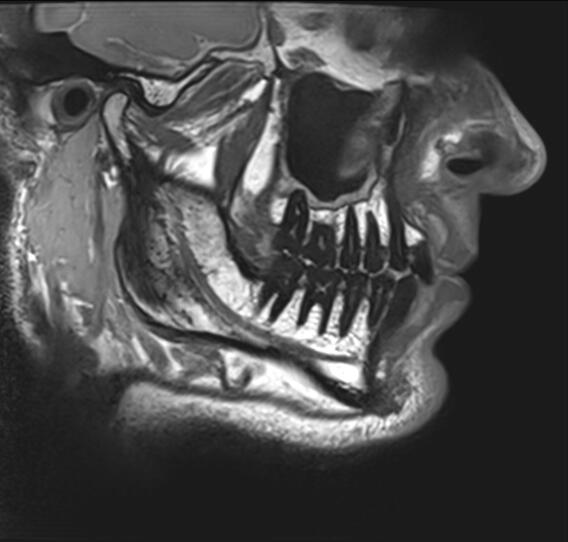
“This image is an adjusted sagittal section of one of our newly-developed 3D proton density anatomy overview sequences,” he said. “The scanning time was 3 minutes 41 seconds total and the image is notable because it shows an unprecedented level of hard and soft tissue detail for this short of a scanning time. Previous versions of a scan like this would have taken perhaps 6-7 minutes or more and offered less image detail.”
The image represents work Gaalaas is doing with the Orthodontic Clinic to determine if such scans can aid in treatment planning. Gaalaas and the orthodontic team are also investigating the role of soft tissue and its role in facial development and orthodontic treatment–something not previously possible in traditional clinical photographs and x-ray scans, which focus on teeth and bones.
“I work in diagnostic radiology and education, so finding improved image quality which will hopefully support improved diagnostic and treatment planning decisions is our whole goal,” reflected Gaalaas, who was quite excited to discover an increase in detail with shorter scan times. “I am excited about this specific sagittal image because it shows detail in a nice variety of tissue types, including the teeth, bone, muscle, fat, nerves and TMJ disk.”
Gaalaas was “thrilled” to win the image contest and extended his gratitude to Donald Nixdorf, DDS, MS, professor and director of the Division of TMD and Orofacial Pain and co-director of the Orofacial MRI Center, as well as Amy Tasca, DDS, PhD, clinical associate professor and Robert J. Isaacson Chair in Orthodontics, Brent Larson, DDS, MS, professor and director of the Division of Orthodontics, and Rob Sellers of Siemens Healthineers, who provided developmental support.
Fahmida Raha, PhD candidate in Molecular, Cellular, Developmental Biology and Genetics (MCDBG) and researcher in Artinger’s lab, submitted a photo of a 22-hour post-fertilization wild-type larval zebrafish embryohead that showed the formation of trigeminal sensory neurons from neural crest cells.
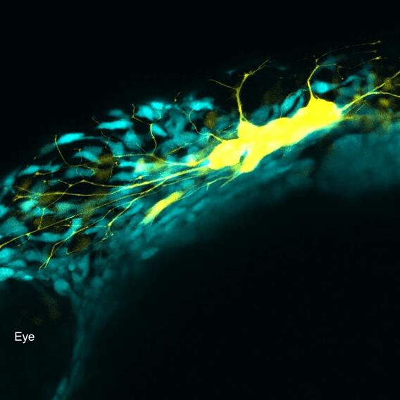
“Sox10+ neural crest cells, shown in cyan, are migrating to complete the formation of the trigeminal ganglia, while trigeminal sensory neurons are marked with HuC, shown in yellow,” she explained” Neurons in the trigeminal ganglia (TG) that innervate the face detect light touch, temperature, pain, and proprioception through skin-projecting. The trigeminal ganglion is formed from neural crest cells; the process starts early in development, around 11-hour post-fertilization. Around 22 hours post-fertilization, the trigeminal neurons start to innervate the face to respond to stimuli and the innervation can be seen with yellow axons. That’s why I love this image, because I tried to capture the first innervation that can be seen with yellow axons here by live imaging confocal microscopy.”
She was excited to participate in the photo contest, which she considered “a good way to express the significance of my research with the help of microscopy.” This image in particular, which shows the moment trigeminal neurons in the embryo extend their axons to innervate the face, is “a beautiful snapshot of how the nervous system begins wiring to sense the world.”
Raha was excited to win the contest and appreciated the opportunity to showcase the importance of visuals in research.
“Visual media helps us actually see what’s happening in research—it turns complex ideas into ,” she reflected. “Images can capture moments, patterns, or processes that words alone can not fully explain, making discoveries easier to understand and beautiful to the eye.”
Both winners’ photographs will be displayed in research conference rooms.