Research Image Contest celebrates the beauty in discovery
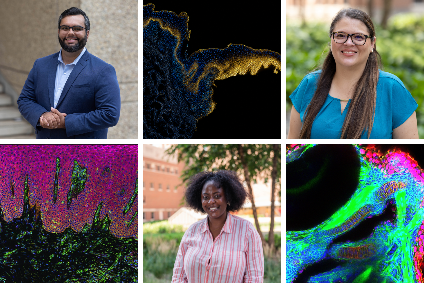
Three School of Dentistry researchers were honored for their commitment to showcasing the beauty of research in this year’s Research Image Contest.
Launched in 2023 and hosted by the Office of Research and Discovery, the Research Image Contest invites researchers to build community by showcasing their research in a visual format.
This year’s winners included Olanrewaju Ajeigbe, PhD candidate, Tomaz Alves da Silva Neto, DDS, MSc, PhD, postdoctoral associate and Silvia Meyer-Nava, PhD, postdoctoral associate.
Olanrewaju Ajeigbe
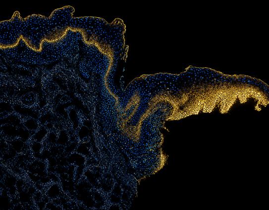
“The picture shows the staining pattern of the protein monocarboxylate transporter-1 (MCT1) across a human oral tissue section. The image shows that as we go from regions of normal epithelium to dysplastic epithelium, MCT1 lights up. This image was the result of transcribing the brown staining usually seen with regular immunohistochemical stains into a golden fluorescence. The golden fluorescence was important because it made a story out of an image that was striking to me as a student of pathology. The beacon-like pattern shows us that this protein can be used to separate danger from safety (just like a beacon) in patients in the clinic. We can use this protein expression to improve early detection of oral cancer in the clinics and hopefully improve prognosis for patients.
This year, in preparation for a talk, I decided to work on one of my slides because I thought there was an important story that could be told there. This image was what I eventually submitted for the contest.
I think science communication is the final frontier for research at the moment, especially considering the current climate we are in as scientists. It is becoming increasingly important to be able to share what you do to the public in simple and clear terms. I think this helps us rally the public around supporting research, and it lets them know why we do what we do. Although good writing and speaking is important to achieve this, as the saying goes, one image can be worth a thousand words. Images can make people feel, and we want others to feel similarly as we do about our science and work.”
Tomaz Alves da Silva Neto
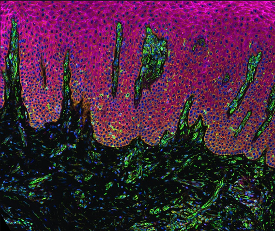
“Captured using Xenium spatial transcriptomics, this image reveals inflamed gingival tissue from a periodontitis subject, spotlighting an understudied interface between the oral epithelium's basal keratinocytes and the underlying stroma. Beyond uncovering transcriptomic signatures during disease, this technology allows us to spatially map and validate these signatures directly within the structural context of oral mucosal barriers. Understanding how immune cell recruitment, host-microbe interactions, and tissue breakdown unfold across these distinct anatomical boundaries is essential to advancing oral biology. Visualizing these spatial relationships at single-cell resolution provides crucial insight into how local microenvironments actively drive or resolve oral inflammation.
Techniques like spatial transcriptomics generate massive amounts of computational data, but they also produce strikingly beautiful visual depictions of living tissue. Powerful imagery translates abstract numbers and complex cellular pathways into an immediate, intuitive narrative. In biomedical research, a compelling image allows scientists and non-experts alike to visualize exact spatial relationships—showing not just what molecules are present, but where they act relative to diseased tissue structures. Beyond communication and education, striking visuals inspire curiosity, making science more accessible and highlighting the natural art embedded within human biology.”
Silvia Meyer-Nava
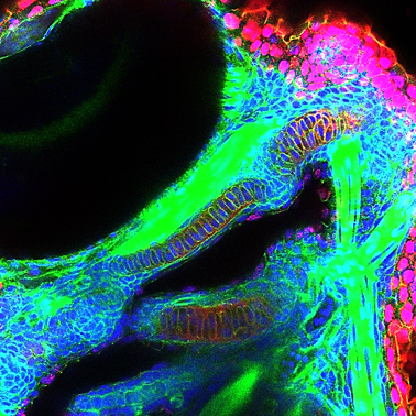
“This confocal image captures a zebrafish embryo at 3 days post-fertilization, revealing the intricate cellular organization of the developing organism. Nuclei are visualized with DAPI in blue, while red highlights the nuclear localization of β-catenin phosphorylated. Phalloidin staining in green marks filamentous actin. The image was taken at 40x and shows the eye and the mandible region. This is important because nuclear β-catenin-pY489 participates in transcriptional regulation and therefore provides information about cell-state/signaling activity.
Much of developmental biology happens at the cellular and molecular level and is invisible to the naked eye. This image provides a glimpse into those fundamental processes, allowing us to visualize how signaling pathways and the cytoskeleton interact during embryonic development.
An image can be very powerful. It can represent beauty while also depicting highly complex biological processes in a way words alone cannot. It can also help bridge the gap between scientists and the general public, making basic research more accessible and inspiring curiosity about how life works.”
--
Winners received a bookstore gift card and the honor of having their image displayed in a research conference room.